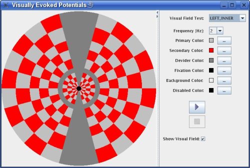
Visually Evoked Potentials

by Puce
Try this:
- Run application using Java Web Start.
Some background information provided by Dr. med. Andreas Gebhardt:
An evoked Potential (EP) test is an electrical potential recorded from a human or animal following presentation of a stimulus and is used to check the condition of the nerve. Evoked potential amplitudes tend to be low, ranging from less than a microvolt to several microvolts, compared to tens of microvolts for EEG (Electroencephalography). Signals can be recorded from cerebral cortex, brainstem, spinal cord and peripheral nerves. Usually the term "evoked potential" is reserved for responses involving either recording from, or stimulation of, central nervous system structures.
Different kinds of evoked potential procedures are used by neurologists: Motor evoked potentials (MEP) test the motor pathways (pyramidal tract) beween the motor cortex and the muscles and are recorded from muscles of upper and lower extremity following direct stimulation of exposed motor cortex Somatosensory evoked potentials (SSEP) test the pathways between the peripheral nerves through the spine to the brain by stimulating nerves with small electrical pulses Visual evoked potentials (VEP) test the visual pathways between the retina (eye) and the brain by presenting alternating checkerboard patterns
Visual evoked potential (VEP) is an evoked potential caused by sensory stimulation of a subject's visual field. Commonly used visual stimuli are flashing lights, or checkerboards on a video screen that flicker between black on white to white on black (invert contrast).
VEPs are very useful in detecting blindness in patients that cannot communicate, such as babies or non-human animals. If repeated stimulation of the visual field causes no changes in EEG potentials, then the subject's brain is probably not receiving any signals from his/her eyes. Other applications include the diagnosis of optic neuritis, which causes the signal to be delayed. Visual evoked potentials are also used in the investigation of basic functions of visual perception. Delayed VEP are a classic finding in optic neuritis (Multiple sclerosis).